Estimated reading time: 11 minutes
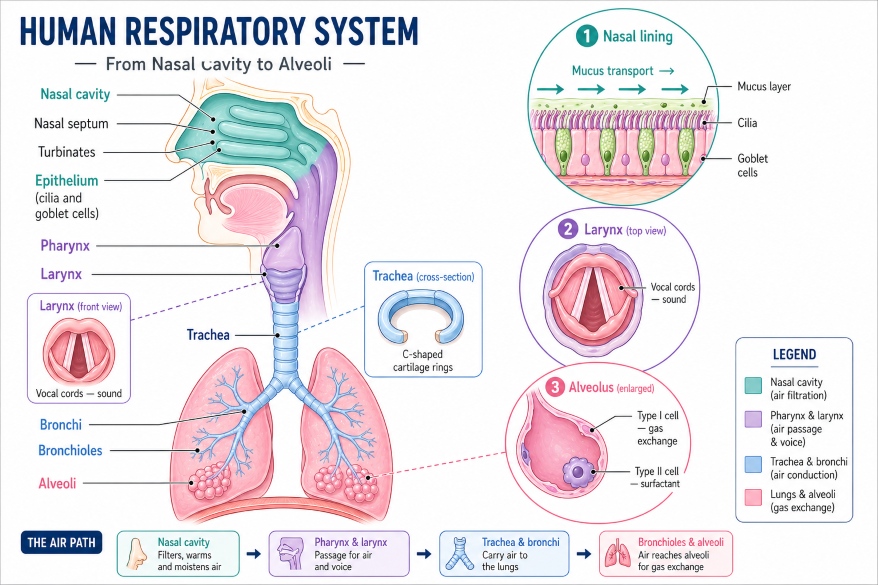
You breathe around 20,000 times every day. Each breath involves many different parts of the human respiratory system working together. At first, most people only think of the lungs. But in reality, the respiratory system has many more components. As a matter of fact, each organ in the respiratory system has a very specific job. Above all, these parts work as a team. In this blog, we take a deep dive into each of the key respiratory system components — what they look like, what they are made of, and what they do.
The Upper Respiratory Tract: Your Body’s First Line of Defence
The upper parts of the human respiratory system include everything from the nostrils down to the larynx. Prior to air reaching the lungs, it must pass through all of these components. At the same time, these organs clean, warm, and moisten the air. As a result, the lungs stay safe and healthy.
The Nose and Nasal Cavity: The Primary Entry Point
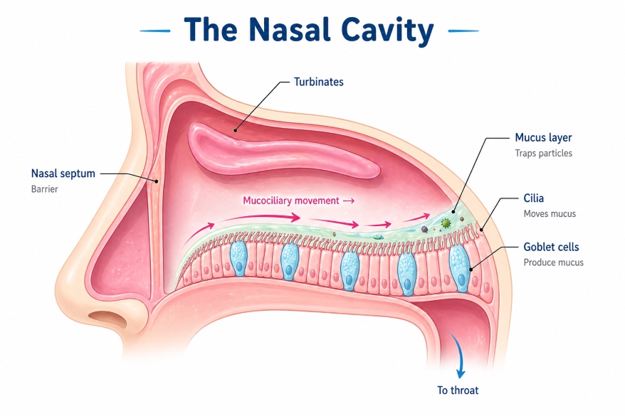
The nasal cavity is where air enters the body. It sits right behind your nose. At this point, the nasal cavity does three key jobs. First, it filters the air. Second, it warms the air. Third, it moistens the air.
So, how does it filter air? To explain, the nasal cavity has a lining full of goblet cells and tiny hairs called cilia. Goblet cells make mucus — a sticky fluid that traps dust, pollen, and germs. After that, the cilia sweep the mucus toward the throat. You then swallow or expel it. This two-step process is called the mucociliary escalator.
What’s more, the nasal cavity contains three bony structures called nasal conchae (or turbinates). These increase the surface area inside the nose. As a result, incoming air spends more time being cleaned and warmed before it moves deeper into the respiratory system. In short, the nasal cavity is far more than just a smell detector. It is the first and most important filter in the human breathing system parts.
The Pharynx: Crossroad in Respiratory System
Below the nasal cavity sits the pharynx, which most people call the throat. It is a muscular tube about 12–14 cm long. The pharynx is one of the shared parts of the human respiratory system — both air and food pass through it.
To enumerate, the pharynx has three sections. First, the nasopharynx sits behind the nasal cavity. Air moves through here. Second, the oropharynx sits behind the mouth. Both food and air pass through here. Third, the laryngopharynx sits at the bottom. Here, food goes toward the food pipe, and air goes toward the larynx.
In addition, the pharynx walls hold lymphatic tissue, including the tonsils and adenoids. These immune tissues watch incoming air for germs. As a result, the pharynx acts as both an air passage and an immune checkpoint.
The Larynx: A Vital Respiratory System Structure
The larynx, also called the voice box, sits just below the pharynx. It is one of the most important respiratory system structures in the upper tract. The larynx protects the airway and produces your voice.
It is made of nine pieces of cartilage. The largest piece is the thyroid cartilage — the bump you can feel on your neck, also called the Adam’s apple.
Inside the larynx sit the vocal cords — two bands of elastic tissue. When air flows up from the lungs, the cords vibrate. These vibrations create sound. After that, your mouth and tongue shape the sound into words.
Above all, the larynx also contains the epiglottis. This is a small, leaf-shaped flap of cartilage. When you swallow, the epiglottis folds down and covers the airway. As a result, food cannot enter the lungs. In like fashion to a trapdoor, it opens for air and closes for food.
The Lower Respiratory Tract: Where Breathing Really Happens
“The lower respiratory tract is an intricate highway — each branch gets smaller and smaller, until air finally reaches the trillions of tiny rooms where life’s most essential exchange takes place.”
The lower parts of the human respiratory system include the trachea, bronchi, bronchioles, alveoli, lungs, and diaphragm. Together, these respiratory system components carry air deep into the body and carry out gas exchange.
The Trachea: A Reinforced Air Pipe
The trachea, also called the windpipe, connects the larynx to the lungs. It is about 10–12 cm long and 2–2.5 cm wide. Without doubt, it is one of the most recognisable parts of the human respiratory system. The trachea stays open at all times. To explain, its walls contain 16–20 C-shaped rings of cartilage along its length. These rings keep the airway from collapsing. At the back of each ring, a band of smooth muscle (the trachealis) bridges the gap. As a result, the oesophagus can expand behind the trachea when you swallow food.
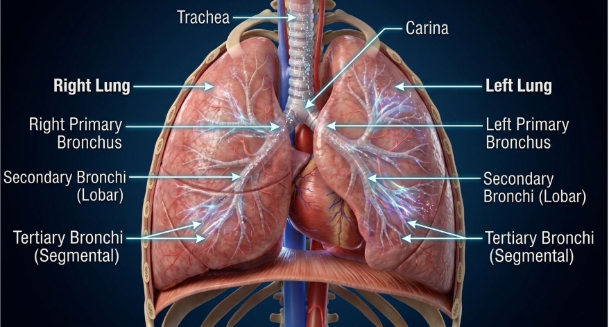
In like fashion to the nasal cavity, the trachea lining also has cilia and goblet cells. These continue the mucociliary escalator, sweeping trapped particles upward and away from the lungs. At the bottom, the trachea splits into two branches at a Y-shaped junction called the carina. These two branches are the left and right bronchi.
The Bronchi: Branch-Like Respiratory System Structures
The bronchi (singular: bronchus) are the two tubes that branch off the trachea. They are among the most important respiratory system structures in the lower tract. One bronchus carries air to the right lung. The other carries air to the left lung. At this point, it is worth noting that the right bronchus is shorter, wider, and more vertical than the left. As a result, if someone accidentally inhales a foreign object, it is more likely to end up in the right lung.
Each bronchus enters its lung at a point called the hilum. After that, it divides into secondary bronchi (one for each lobe) and then into tertiary bronchi (one for each lung segment). In short, the bronchi spread air to every part of both lungs. In similar fashion to the trachea, the bronchi also have cartilage, smooth muscle, cilia, and mucus in their walls. So they continue both the structural support and the cleaning work of the upper respiratory system components.
The Bronchioles: The Tiniest Airways
As the bronchi keep dividing, they get smaller and smaller. At last, they lose their cartilage walls and become bronchioles — tubes less than 1 mm wide. These are among the finest components of the human respiratory system. Unlike the bronchi, bronchioles have no cartilage. Instead, their walls contain smooth muscle wrapped in elastic fibres. This muscle can tighten or relax to control airflow. For instance, during exercise, bronchioles widen to let more air in. Sooner or later, after exercise, they relax back to normal size.
To point out a common problem — in asthma, the smooth muscle of the bronchioles tightens abnormally. This narrows the airway and makes breathing hard. This condition is called bronchoconstriction. As a result, people with asthma use inhalers to relax the bronchiole muscles and open the airway. In due time, the bronchioles narrow into terminal bronchioles. These lead to the final and most important part of the respiratory system — the alveoli.
The Alveoli: Where Life Happens
The alveoli (singular: alveolus) are tiny, balloon-shaped air sacs at the very end of the bronchioles. Of all the parts of the human respiratory system, the alveoli are the most critical. This is where gas exchange actually happens.
Each human lung holds about 300–500 million alveoli. Together, they give the lungs a total surface area of roughly 70 m². To illustrate — that is as large as a tennis court! As a result, your body can exchange a huge amount of gas very quickly.
Each alveolus has two types of cells:
- Type I alveolar cells — these are extremely thin and flat. They cover about 95% of the alveolar surface. Their thinness allows oxygen and CO₂ to pass through quickly and easily.
- Type II alveolar cells — these are smaller and rounder. They make surfactant — a substance that stops the alveolus from collapsing when you breathe out. Without surfactant, your air sacs would stick shut.
Each alveolus sits inside a mesh of capillaries (tiny blood vessels). Oxygen crosses from the alveolus into the blood through the ultra-thin blood-air barrier — just 0.5–1 micrometre thick. At the same time, carbon dioxide crosses in the opposite direction. After that, you exhale the CO₂ out of your body.
In short, the alveolus is where breathing becomes biology. It is, without doubt, the most vital of all the human breathing system parts.
The Lungs: The The Central Organs of the Respiratory System
The lungs are the two large organs that house all the bronchi, bronchioles, and alveoli. They sit in the thoracic cavity (chest), protected by the rib cage and separated from each other by a space called the mediastinum (which contains the heart and major blood vessels). The right lung is larger, with three lobes — the upper, middle, and lower lobe. The left lung has only two lobes — the upper and lower — because the heart occupies the space on the left side.
Each lung is covered by a double-layered membrane called the pleura. The outer layer (parietal pleura) lines the inside of the chest wall. The inner layer (visceral pleura) covers the surface of the lung. Between the two layers is a thin space called the pleural cavity, which contains a small amount of pleural fluid. This fluid acts as a lubricant, allowing the lungs to expand and contract smoothly with each breath.
The Diaphragm: The Engine of the Respiratory System Parts
Below the lungs sits the diaphragm — a dome-shaped sheet of skeletal muscle that is the primary muscle of breathing. It separates the thoracic cavity from the abdominal cavity. The diaphragm is supplied by the phrenic nerve, which originates from the C3, C4, and C5 nerve roots in the neck (remembered by the medical phrase: “C3, 4, and 5 keep the diaphragm alive”). When the phrenic nerve fires, the diaphragm contracts and flattens downward, increasing the volume of the chest cavity and drawing air into the lungs (inhalation). When it relaxes, it curves back upward, decreasing chest volume and pushing air out (exhalation).
During normal quiet breathing, the diaphragm does about 70–80% of the breathing work. During deep breathing or exercise, additional muscles, including the intercostal muscles between the ribs and the accessory muscles in the neck and abdomen — assist the diaphragm.
Table 1: Respiratory System: Structure & Function
| Part | Location | Key Structure | Main Function |
|---|---|---|---|
| Nasal cavity | Behind the nose | Cilia, goblet cells, turbinates | Filter, warm, moisten air |
| Pharynx | Throat | Lymphatic tissue, tonsils | Air and food passage; immune defence |
| Larynx | Top of trachea | Vocal cords, epiglottis, cartilage | Voice production; airway protection |
| Trachea | Neck/upper chest | C-shaped cartilage rings | Air conduction |
| Bronchi | Inside lungs | Cartilage, smooth muscle, cilia | Air distribution to lung lobes |
| Bronchioles | Deep in lungs | Smooth muscle, elastic fibres | Fine airflow control; no cartilage |
| Alveoli | Lung tips | Type I & II cells, capillaries | Gas exchange (O₂ in, CO₂ out) |
| Lungs | Thoracic cavity | Lobes, pleura, blood vessels | House all gas exchange structures |
| Diaphragm | Below lungs | Skeletal muscle, phrenic nerve | Primary breathing muscle |
Summary: Parts of Human Respiratory System
To sum up, each part of the human respiratory system plays a very specific and important role:
- The nasal cavity filters, warms, and moistens air using cilia, goblet cells, and turbinates.
- The pharynx is a shared passage for air and food, with immune tissues (tonsils) for protection.
- The larynx protects the airway with the epiglottis and enables speech through the vocal cords.
- The trachea is reinforced with C-shaped cartilage rings and uses mucociliary clearance to clean the airway.
- The bronchi branch into left and right lungs, further dividing into lobar and segmental bronchi.
- The bronchioles are tiny, cartilage-free airways controlled by smooth muscle; they are affected in asthma.
- The alveoli are the site of gas exchange, lined by ultra-thin Type I cells and surfactant-producing Type II cells.
- The pleura lubricates the lungs for smooth expansion.
- The diaphragm is the main breathing muscle, controlled by the phrenic nerve.
In conclusion, understanding the parts of the human respiratory system at this level gives you a strong foundation for all of biology and medicine. Sooner or later, every system in the body connects back to how well we breathe. You are building knowledge that will serve you for years to come — keep going!
Frequently Asked Questions (FAQs)
The mucociliary escalator is a defence system in the respiratory system parts. Goblet cells make mucus that traps dust and germs. Cilia then sweep the mucus upward toward the throat. This keeps the lungs clean and safe.
Bronchi are larger airways with cartilage walls. Bronchioles are smaller, cartilage-free airways with smooth muscle walls. The bronchioles lead air all the way to the alveoli.
Type I cells are very thin and flat — they cover most of the alveolar surface and allow fast gas diffusion. Type II cells are smaller and produce surfactant, which stops the alveoli from collapsing during exhalation.
The phrenic nerve — arising from neck vertebrae C3, C4, and C5 — controls the diaphragm. If this nerve gets damaged, the diaphragm stops working and breathing becomes very difficult.
The blood-air barrier is the ultra-thin wall between the air inside an alveolus and the blood inside a capillary. It is only 0.5–1 micrometre thick. Oxygen and carbon dioxide cross this barrier during gas exchange.
Reference
Rogers, T. D., Button, B., Kelada, S. N. P., Ostrowski, L. E., Livraghi-Butrico, A., Gutay, M. I., Esther, C. R., & Grubb, B. R. (2022). Regional differences in mucociliary clearance in the upper and lower airways. Frontiers in Physiology, 13, 842592. https://doi.org/10.3389/fphys.2022.842592
Knudsen, L., & Ochs, M. (2018). The micromechanics of lung alveoli: Structure and function of surfactant and tissue components. Histochemistry and Cell Biology, 150(6), 661–676. https://doi.org/10.1007/s00418-018-1747-9
- About the Author
- Latest Posts

Ayushi Shukla is a biotechnologist and science communicator specializing in the intersection of genomic data and public health. Previously, she served as a Core Member and R&D Lead at the HealthTech venture Eat Wisely Bro, where she spearheaded research initiatives and translated emerging medical data into product strategy. She holds an M.Sc. in Biotechnology from the TERI School of Advanced Studies, where her research at The Energy and Resources Institute (TERI), New Delhi, focused on Environmental Biotechnology and Bioremediation.
As a Research Scholar at TERI, Ayushi developed a plant growth-promoting bacterial consortium for high-salinity agricultural conditions, managing daily laboratory procedures and molecular workflows. Her technical portfolio on GitHub demonstrates her dry lab expertise, featuring end-to-end bioinfomatics pipelines for RNA-Seq analysis, Phylogenetic modeling, and a functional prototype for Mycobacterium tuberculosis Genomic Surveillance & Dashboard.
Her clinical foundation includes advanced training in Somatic NGS and Precision Oncology from the Cancer Research Centre at Tata Memorial Centre, and a professional certification in Good Clinical Practice (GLP) from the NIDA Clinical Trials Network. Ayushi bridges the gap between raw genomic data and student-friendly narratives to help the next generation of scientists understand the transformative power of modern biology.