The human eye is an important sense organ that helps us see colors, shapes, light, and movement. It also helps us enjoy the beautiful world around us. Light enters the eye through the clear front part called the cornea. The iris controls the size of the pupil to manage how much light enters the eye. A convex lens then focuses the light onto the retina at the back of the eye.
The retina contains special cells called rods and cones. Rods help us see in dim light, while cones help us see colors and bright light. The optic nerve carries messages from the retina to the brain, which helps us see images clearly. In this article, you will learn how the human eye works and how its different parts help us see properly.
Key Takeaways
- The cornea helps focus light.
- The iris controls the amount of light entering the eye.
- The pupil allows light to enter the eye.
- The lens focuses light on the retina.
- The retina converts light into signals for the brain.
- The optic nerve carries these signals to the brain.
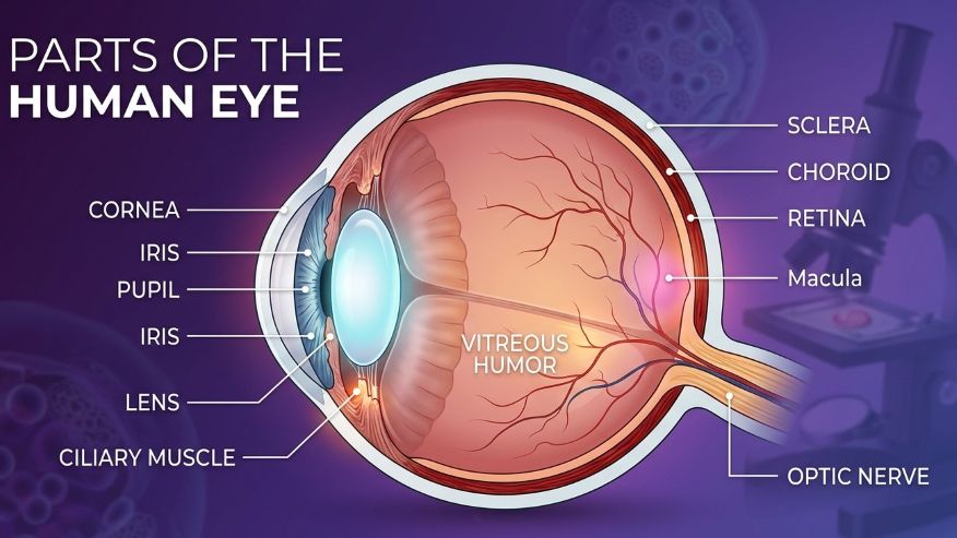
Structure of the Human Eye
The human eye is around 2.3 cm in diameter and resembles a little ball with an almost spherical shape. The vitreous humor is a jelly-like material found inside the eye. This jelly supports the inside components of the eye and aids in maintaining its shape.
The eye is made up of numerous vital components that cooperate to enable excellent vision. The cornea, iris, pupil, lens, retina, and optic nerve are a few of these components:
Here is a brief of the parts of the Human eye:
- Sclera – The sclera is the white outer covering of the eye. It protects the eye.
- Cornea – The cornea is the clear front part of the eye. Light enters the eye through the cornea.
- Iris – The iris is the colored part of the eye. It controls how much light enters the eye.
- Pupil – The pupil is the small black opening in the center of the eye. It lets light enter the eye.
- Lens – The lens helps focus light on the retina. It changes shape to help us see near and far objects clearly.
- Retina – The retina is a light-sensitive layer at the back of the eye. It sends messages to the brain.
- Optic Nerve – The optic nerve carries messages from the eye to the brain.
Special Cells in the Retina
- Cones aid in color and intense light perception.
- Rods enable us to see at night and in low light.
- Blind Spot: The blind spot is a little region devoid of nerve cells, making it impossible to see an image.
The eye has six muscles called eye muscles. These muscles help the eye move in different directions.
Cornea: The Transparent Front Layer
The transparent, curving front portion of the eye is called the cornea. The cornea allows light to enter the eye. It facilitates clear vision by bending and focusing light. Before light reaches the lens and retina, it is mostly focused by the cornea.
Additionally, the cornea shields the eye from bacteria, dust, and damaging UV radiation from the sun. Tears help maintain the cornea’s health by keeping it clean and moist.
The cornea has five main layers:
- Epithelium – The outer layer that protects the eye and absorbs oxygen from tears.
- Bowman’s Membrane – A thin protective layer made of collagen.
- Stroma – The thickest layer that helps keep the cornea round and clear.
- Descemet’s Membrane – A strong layer that protects the inner parts of the eye.
- Endothelium – The inner layer that removes extra fluid and keeps the cornea clear.
All these layers work together to help us see properly and protect the eye.
Iris: The Colored Part of the Eye
The colored portion of the eye is called the iris. It regulates both the pupil’s size and the amount of light that enters the eye. The pupil gets smaller in bright light and larger in dim light. A healthy iris shields the eye from excessive light and aids in good vision. Injuries, illnesses, surgeries, and congenital defects can occasionally harm the iris. This may result in changes to the look of the eye, glare, or blurry vision.
The iris is crucial because it protects the eye and facilitates comfortable vision.
Pupil: The Opening That Lets Light In
The pupil is the small black opening in the center of the eye. It lets light enter the eye and reach the retina.
The pupil changes size depending on the light:
- In bright light, the pupil becomes smaller.
- In dim light, the pupil becomes bigger.
The pupil helps control how much light enters the eye and helps us see clearly. It also allows fluid to move inside the eye.
The iris muscles control the size of the pupil:
- The sphincter muscle makes the pupil smaller.
- The dilator muscle makes the pupil bigger.
Lens: The Part That Focuses Light
- To help us view both close and distant objects, the lens undergoes a morphological change.
- To see distant objects clearly, it gets thinner.
- To clearly see objects in the vicinity, it gets thicker.
This ability is called accommodation.
What the Lens is Made Of ?
Water and unique proteins known as crystallins make up the majority of the lens. These maintain the lens’s transparency and clarity.
Lens Growth
The lens keeps growing slowly throughout life as new cells are added.
Common Lens Problems
- Cataract – The lens becomes cloudy and makes vision blurry.
- Presbyopia – As people get older, the lens becomes less flexible, making it harder to see nearby objects clearly.
Retina: The Light-Sensitive Layer
The retina is the light-sensitive layer at the back of the eye. It helps us see by changing light into messages for the brain.
The retina has special nerve cells that work together:
- Photoreceptors – These cells detect light.
- Connecting nerve cells – These cells pass messages from the photoreceptors to other cells.
- Ganglion cells – These cells send messages to the brain through the optic nerve.
- Horizontal cells and amacrine cells – These cells help the eye process and organize visual information.
Light enters the eye and reaches the retina. The retina changes the light into electrical signals. These signals travel through the optic nerve to the brain. The brain then helps us understand what we see.
The retina has many connected cells that help us see shapes, colors, brightness, and movement clearly.
Optic Nerve: The Connection Between Eye and Brain
The optic nerve is an important nerve that connects the eye to the brain. It carries visual messages from the retina to the brain so we can see and understand images. The optic nerve is made of millions of tiny nerve fibers that work together to send signals. These signals travel quickly from the eye to the brain.
The optic nerves from both eyes meet at a place called the optic chiasm. Here, some nerve fibers cross over to the other side of the brain. This helps the brain combine information from both eyes and helps us see clearly.
The optic nerve also helps control:
- The pupil’s reaction to light
- The eye’s ability to focus on objects
A small area called the blind spot is found where the optic nerve leaves the eye. This area cannot detect images because there are no light-sensitive cells there.
Sclera: The White Protective Layer
The sclera is the white outer part of the eye. It is strong and helps protect the eye. The sclera also helps the eye keep its round shape. The sclera blocks extra light from entering the eye from the sides. This helps us see clearly. The sclera covers most of the eyeball. It joins with the cornea at the front of the eye and connects to the optic nerve at the back.
Layers Around the Sclera
- Tenon’s Capsule – A thin layer that helps the eye move smoothly.
- Episclera – A protective layer around the sclera.
- Stroma – The thickest layer that keeps the eye strong.
Functions of the Sclera
- Protects the eye
- Helps the eye keep its shape
- Blocks extra light
- Helps the eye muscles move the eye
The sclera is important because it keeps the eye safe and strong.
Important Parts Inside the Sclera
- Collagen – Collagen is a strong protein that gives the sclera strength and helps the eye keep its shape.
- Proteoglycans – These substances help hold water inside the sclera and keep the eye firm and healthy.
- Elastic Fibers –These fibers help the sclera stretch a little and return to its normal shape.
- Fibroblasts – These are special cells that help repair and build the sclera when the eye is injured.
Functions of These Parts
- Helps keeping the eye strong
- Helps the eye keeping its round shape
- Protect the inner parts of the eye
- Help repair damaged eye tissues
- Support clear vision
All these parts work together to keep the eye healthy, strong, and protected.
Conjunctiva: The Protective Covering
The conjunctiva is a thin, clear layer that covers the white part of the eye and the inside of the eyelids. It helps protect the eye, keeps it moist, and helps the eyes move smoothly. The conjunctiva also makes tears and mucus to keep the eye wet and protects the eye from germs and dust.
Aqueous Humor and Vitreous Humor
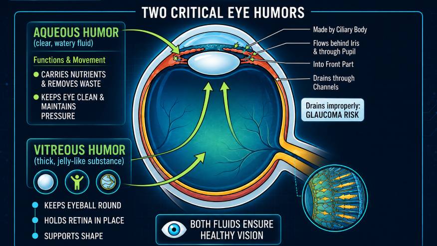
The eye has two important fluids called the aqueous and the vitreous humor.
The aqueous humor is a clear, watery fluid found in front of the lens. The vitreous humor is a thick, jelly-like substance found behind the lens and in front of the retina.
Functions of Aqueous Humor
The aqueous humor helps:
- Carries oxygen and nutrients to the eye
- Removes waste from the eye
- Keeps the eye clean and healthy
- Maintain the shape and pressure of the eye
This fluid is always being made and drained from the eye to keep the pressure balanced.
How Aqueous Humor Moves
The fluid is made by the ciliary body inside the eye. It flows:
- Behind the iris
- Through the pupil
- Into the front part of the eye
- Out through tiny drainage channels
If the fluid does not drain properly, it can cause a disease called glaucoma.
Vitreous Humor
The vitreous humor is a jelly-like substance that fills the back part of the eye. It helps:
- Keep the eyeball round
- Hold the retina in place
- Support the shape of the eye
Both the aqueous and vitreous humor are important for keeping the eyes healthy and helping us see clearly.
Rods and Cones: Special Cells in the Retina
Rods are special cells found in the retina at the back of the eye. They help us see in dim light and at night. Rods help us see black, white, and shades of grey. They also help us notice shapes, shadows, and movement. Rods cannot see colours. These cells are mostly found around the outer part of the retina, so they also help with side vision (peripheral vision). Rods are very sensitive to light, which helps us see even in dark places.
The retina at the rear of the eye has unique cells called cone cells. They enable us to see colors and clear details. Bright light, such as sunlight or a bright room, is ideal for cone cells. We can see colors like red, green, and blue thanks to these cells. They also help with reading, drawing, and clear vision. The macula, the central region of the retina, contains most of the cone cells. The fovea, a small region in the center, helps us see the sharpest and clearest images.
Interesting Facts About the Human Eye Structure:
The Cornea Can Breathe From the Air
The cornea is the only part of the human body that does not have blood vessels. It gets oxygen directly from the air.
The Retina Works Like a Powerful Camera
The retina has millions of light-sensitive cells that help us see clear and detailed images.
Images Are Formed Upside Down in the Eye
The lens focuses images upside down on the retina. The brain then turns the image the correct way so we can see properly.
The Iris Moves Very Quickly
The iris muscles are some of the fastest muscles in the human body. They quickly make the pupil bigger or smaller to control the amount of light entering the eye.
Conclusion
An incredible sense organ is the human eye. To see colors, shapes, light, and movement clearly, the eye’s various components work together. Each component of the eye, including the cornea, iris, lens, retina, optic nerve, and others, has a specific function.
We canread, learn, move safely, and appreciate the world around us when our eyes are healthy. Maintaining strong and healthy eyesight requires proper eye care.
Frequently asked questions (FAQs)
The human eye is a sense organ that allows humans to perceive color, form, light, and movement.
The iris regulates the amount of light that enters the eyes by altering the pupil’s size.
The cornea shields the eye and facilitates light entry.
Light is converted into messages by the retina and sent to the brain.
Cones enable humans to discern color details and clarity, whereas rods detect weak light.
References:
- Boote, C., Sigal, I. A., Grytz, R., Hua, Y., Nguyen, T. D., & Girard, M. J. A. (2020). Scleral structure and biomechanics. Progress in Retinal and Eye Research, 74, 100773. https://doi.org/10.1016/j.preteyeres.2019.100773
- Ferro Desideri, L., Arun, K., Doherty, G., Bernardi, E., & Anguita, R. (2024). Iris reconstruction: A surgeon’s guide. Journal of Clinical Medicine, 13(9), 2706. https://doi.org/10.3390/jcm13092706
- London, A., Benhar, I., & Schwartz, M. (2012). The retina as a window to the brain—from eye research to CNS disorders. Nature Reviews Neurology, 9(1), 44–53. https://doi.org/10.1038/nrneurol.2012.227
- Tomy, R. (2019). Pupil: Assessment and diagnosis. Kerala Journal of Ophthalmology, 31(2), 167. https://doi.org/10.4103/kjo.kjo_48_19
- About the Author
- Latest Posts

Ms. Adrita Roy is a dual master’s holder specializing in Biological Sciences and Sustainable Development. She completed her Master’s in Biological Sciences from Bangalore University in 2025, followed by a Master’s in Sustainable Development from the University of Sussex in 2026.
With over three years of professional experience across biosciences, sustainability, education, ESG consulting, and science communication, Ms. Roy has developed a multidisciplinary profile focused on research, sustainability, and impactful communication.
Her subject expertise includes Biological Sciences, Sustainable Development, ESG consulting and sustainability frameworks, science communication, environmental awareness communication, and sustainability-focused content development. She also possesses expertise in Biology and Chemistry academic content development for Classes 11 and 12, including MCQs, assignments, case studies, and educational learning resources.
Ms. Roy is experienced in scientific writing, journal drafting, research communication, policy drafting, stakeholder engagement, sustainability reporting, web content creation, marketing communication, case study preparation, and master sheet preparation. Her multidisciplinary background enables her to combine research-oriented knowledge with effective communication strategies in the fields of science, sustainability, and education.